
Internationally, University of Antwerp focuses on several strategic research areas. One of the key domains is electron microscopy for materials science. The EMAT research group is a global leader in this field, with a particular focus on electron tomography using transmission electron microscopy (TEM).
Professor Dr Sara Bals, renowned for her work in electron tomography and its application in the study of nanomaterials such as perovskite crystals, serves as spokesperson of the group.
From 2D to 3D atomic imaging
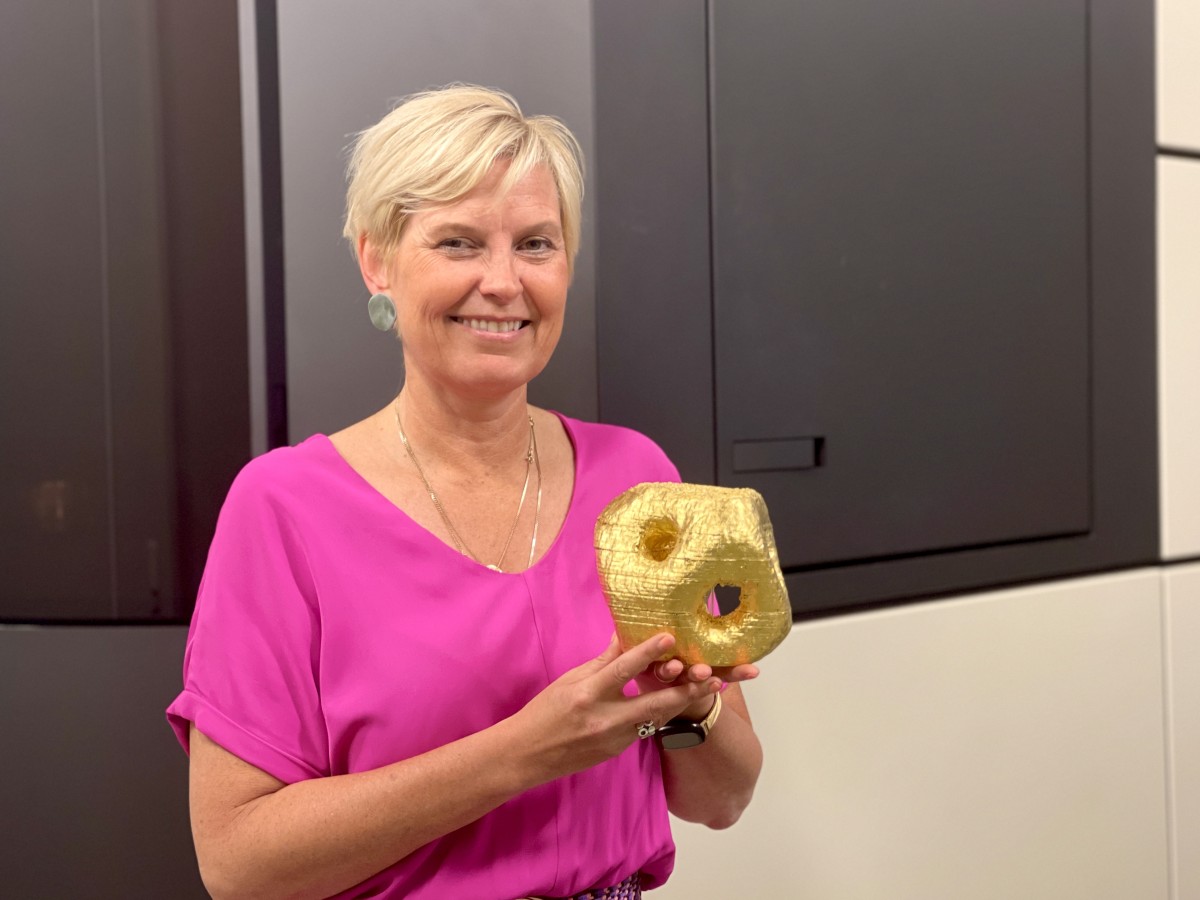
She has played a central role in transforming transmission electron microscopy (TEM) from a two-dimensional imaging technique into a fully three-dimensional (3D) measurement method capable of determining the positions of individual atoms in nanomaterials. The insights obtained through this approach are of major technological importance, as the properties of modern nanomaterials are largely determined by their three-dimensional structure.
Like optical microscopy, an electron microscope produces a projection image—a two-dimensional shadow of a three-dimensional object. “For 3D imaging, we use the same principle as a medical CT scanner,” Professor Bals explains. “The detector rotates around the patient, taking images from different angles, which are then combined using mathematical algorithms into a 3D reconstruction. In our case, the nanoparticles are tilted on their support inside the microscope.”
Imaging under real-world conditions
The innovative and pioneering nature of Professor Bals’ research was recognised in 2013 with the award of an ERC Starting Grant (Colouratom) for the project Colouring Atoms in 3 Dimensions. In 2019, she received an ERC Consolidator Grant for 3D Structure of Nanomaterials under Realistic Conditions (Realnano).
By combining state-of-the-art electron microscopy with advanced 3D reconstruction algorithms, her group has made it possible to routinely determine both the positions and chemical identity of individual atoms in a wide range of nanomaterials. Today, these measurements can also be performed under realistic operating conditions, including heating, liquid environments, and gas-flow experiments. This allows researchers to study nanomaterials while they are functioning.
This capability is particularly important for the nanoscale characterisation of beam-sensitive perovskite materials under realistic working conditions.
“Traditionally, samples are examined in a vacuum environment, where they behave differently from how they do under operational conditions,” Professor Bals explains. “We create a much more relevant environment for our observations.”
Both ERC projects were followed by Proof of Concept (PoC) projects, aimed at bringing the resulting innovations to industry. HYPERGRAPH developed a method for producing high-quality graphene grids with reliable performance and high production yields. SEEBIC – still ongoing – extends the functionality of standard TEM setups for 3D imaging of nanomaterials.
Advanced instrumentation
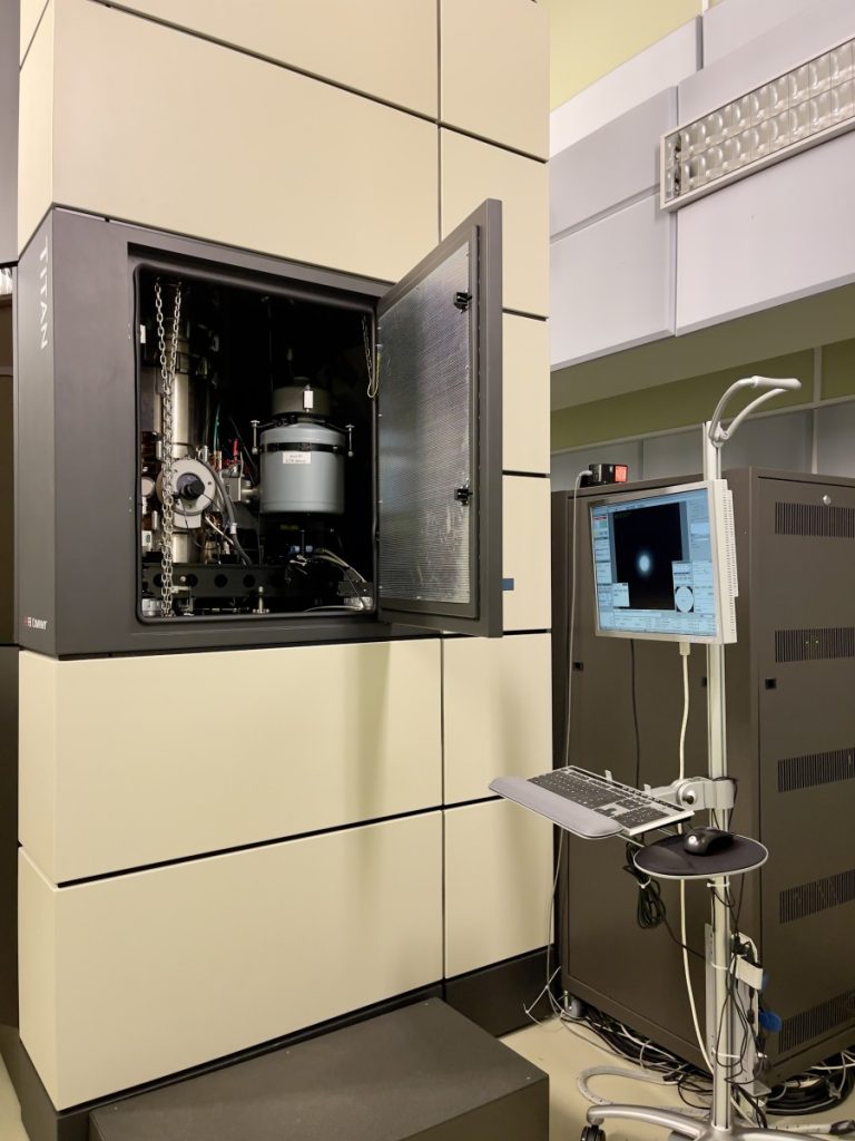
EMAT operates several state-of-the-art transmission electron microscopes, as well as a range of conventional yet highly versatile instruments. In total, the facility includes six transmission electron microscopes, four of which are aberration-corrected. One system even features double aberration correction, enabling a resolution of 0.05 nm—ideal for mapping atomic positions of both heavy and light elements.
This level of instrumentation remains rare in Europe. The University of Antwerp was among the first European institutions to implement such technology. EMAT also operates three scanning electron microscopes (SEMs).
Important contributions
“Our laboratory has made an important contribution to improving image resolution,” says Professor Bals. “Traditionally, this requires highly energetic electrons, but that increases the risk of damaging sensitive samples. To avoid this, we use lower accelerating voltages. In addition, our reconstruction algorithms incorporate prior knowledge about the material—for example, that it consists of identical atoms—which enables high-quality 3D reconstructions from fewer images.”
Samples are mounted on copper grids coated with an ultrathin layer of graphene. “The quality of commercially available graphene grids is often disappointing. We therefore developed our own method for producing higher-quality graphene supports, which we have patented.”
People and research community
Today, the EMAT research team consists of six professors. Under the supervision of Professor Bals, thirty PhD students have completed their doctorates, three of whom received awards from the Belgian Society for Microscopy. The laboratory currently hosts nearly 25 PhD researchers and a similar number of postdoctoral researchers.
NANOlight
Within the University of Antwerp, EMAT collaborates with other research groups through the NANOlight centre of excellence. This consortium consists of six research groups with complementary expertise in nanoscience and nanomaterials, all based at the University of Antwerp. It brings together physics (EMAT, Sara Bals and Sandra Van Aert and COMMIT, Milorad Milosevic), chemistry (AXIS, Koen Janssens), bio-science engineering (A-PECS – Karolien De Wael, A-PECS – Sammy Verbruggen), and economics (EnvEcon, Steven Van Passel).
The NANOlight teams aim to rationally design and exploit nanomaterials and their interaction with light in fields such as plasmonics, electrosensing, and photocatalysis, as well as applications in art conservation, environmental science, and sustainable energy.
Science meets cultural heritage: The Scream
EMAT’s research also extends to the humanities and cultural heritage.
For example, supporting Professor Koen Janssens’ AXIS team within Nanolight, the group investigates the composition and degradation of pigments in historical paintings.
One of the most famous works analysed is The Scream (1893) by Edvard Munch, where researchers identified and visualised the degradation of the cadmium yellow (CdS) pigment.

Collaboration and applications
EMAT also collaborates extensively with national and international research institutions and industry partners. “In such projects, we focus on structural characterization, while other partners handle nanoparticle synthesis and practical applications. For example, we can visualise the morphology of peptide fibres decorated with Au nanoprticles. This information may help medical specialists in the future to determine the optimal wavelength of light to destroy ‘bad’ peptides while leaving ‘good’ ones unaffected.”
Chirality and nanomedicine
A key ongoing project involving EMAT is Chiral-Pro. Chirality—the property of an object lacking mirror symmetry—is found in both biological and synthetic materials. While advances in chiral inorganic nanoparticles have demonstrated strong potential, their tailored design for biological interactions remains challenging.
Much of the research in this field has focused on chiral nanostructures formed through directed self-assembly of gold nanorods on chiral templates. More recent approaches use seeded-growth strategies to introduce chiral features in colloidal nanoparticles in the presence of molecular chiral inducers. These concepts open up broad possibilities by varying inducers, co-surfactants, seed morphologies, and metal compositions.
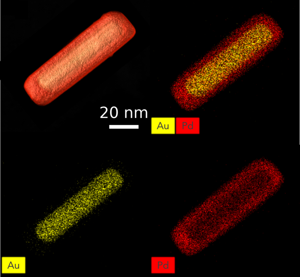
The ERC-funded project aims to develop methodologies for engineering chiral nanoparticles with exceptional optical activity, enabling precise enantioselective interactions with biological structures such as proteins and amyloid fibrils.
“By uncovering the role of chirality in molecular recognition, we aim to enable the programmed assembly of optically active nanofibres and explore applications in biosensing and metamaterials,” says Professor Bals.
“We also aim to use AI tools to predict optimal nanoparticle morphologies for selective and strong binding with proteins. This could lead to significant advances in disease detection, including cancer and Alzheimer’s disease.”
Future health applications
“In addition to fundamental scientific breakthroughs, this research may provide a toolbox for moving from trial-and-error approaches to the rational design of chiral nanostructures with predefined properties.” Chiral-Pro is expected to accelerate the development of nanoparticles for applications such as amyloid detection, inhibition of fibrillation, metamaterials, contrast agents, nanoscale adjuvants, enzyme mimics, and antimicrobial agents.
International collaboration
The Chiral-Pro consortium brings together experts in colloidal synthesis, predictive modelling of nanoparticle–protein interactions, and advanced electron microscopy from Belgium, Spain, and the United States. Partners include EMAT, the BioNanoPlasmonics group at CIC biomaGUNE, the Biomimetic Materials group at CINBIO (University of Vigo), and the Chemical Engineering group of Professor Nicholas A. Kotov at the University of Michigan.
The Vigo laboratories focus on nanoparticle synthesis and growth mechanisms. EMAT develops fast electron tomography techniques for high-throughput 3D characterisation of nanoparticles. The Michigan group contributes expertise in artificial intelligence for predicting interactions between nanoparticles and biomolecules.
Electron microscopy for Materials Science (EMAT)
Universiteit Antwerpen
Campus Groenenborger
Groenenborgerlaan 171
B-2020 Antwerpen
T: +32 32 65 32 48
:+To+see+what+no+one+saw+before&description=&image=https://belgian-research.eu/wp-content/uploads/sara-bals-1024x768.jpeg)